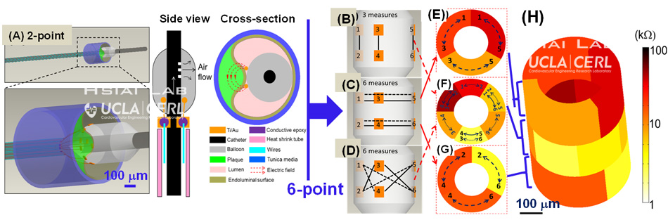
Intravascular deployment of the integrated EIS sensor And IVUS transducer to assess lipid-laden plaques

Figure . (A) Conceptual scheme depicts the deployment of the integrated sensor consisting of an EIS sensor and an IVUS transducer to assess lipid-rich plaques. The IVUS sensor visualizes the aorta lumen, and the imaging information provides guidance for EIS characterization of the plaques by aligning the EIS sensor (2-point electrode) at the plaque. PBS: Phosphate-buffered saline solution. (B) The design of the integrated sensor highlights the mechanism for IVUS-guided EIS measurement. The IVUS transducer is positioned inside the inner catheter (ID: 1 mm, OD: 1.3 mm) with an imaging window of 2 cm to 10 cm. The EIS sensor is affixed to the balloon, which is anchored to the outer catheter (ID: 1.7 mm, OD: 2 mm). External pump generates air pressure to inflate or deflate the balloon, ranging from 2.3 mm to 6 mm in diameter.
Ultrasonic Transducer-Guided Electrochemical Impedance Spectroscopy to Assess Lipid-Laden Plaques.
Ma J, Luo Y, Sevag Packard RR, Ma T, Ding Y, Abiri P, Tai YC, Zhou Q, Shung KK, Li R, Hsiai T.
Sens Actuators B Chem. 2016 Nov 1;235:154-161. Epub 2016 May 7.
PMID: 27773967
Design of matching layers for high-frequency ultrasonic transducers.
Fei C, Ma J, Chiu CT, Williams JA, Fong W, Chen Z, Zhu B, Xiong R, Shi J, Hsiai TK, Shung KK, Zhou Q.
Appl Phys Lett. 2015 Sep 21;107(12):123505. Epub 2015 Sep 24.
A Review of Intravascular Ultrasound-based Multimodal Intravascular Imaging: The Synergistic Approach to Characterizing Vulnerable Plaques.
Ma T, Zhou B, Hsiai TK, Shung KK.Ultrason Imaging. 2016 Sep;38(5):314-31. doi: 10.1177/0161734615604829. Epub 2015 Sep 22. Review.